 |
| Fig1: Atrial Septal Defect |
 |
| Fig 2: Atrial Septal Defect. |
Eighty-five per cent of ASDs are of the secundum type, 11% of the primum type and 4% sinus venosus defects involving the inferior vena cava (IVC) or superior vena cava (SVC).
 |
| Fig 3: SVASD = Sinus Venosus Atrial Septal Defect; OSASD = Ostium Secundum Atrial Septal Defect; OPASD = Ostium Primum Atrial Septal Defect. |
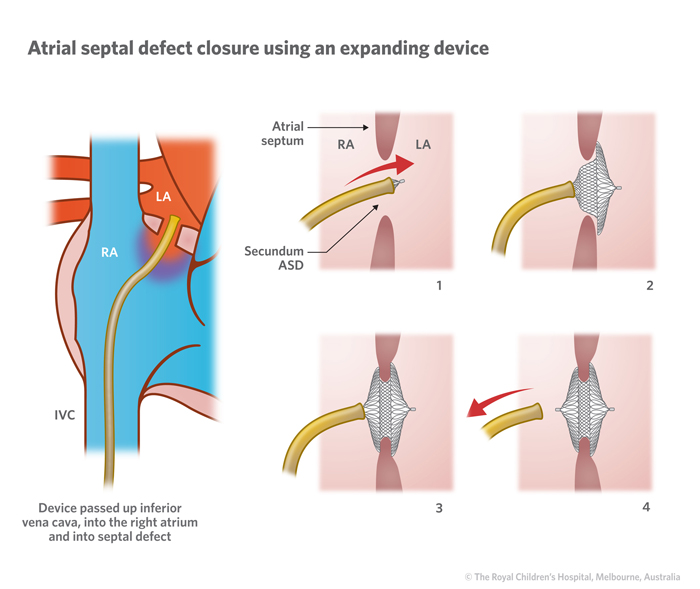
Many ASDs are now being closed using percutaneous closure devices.
 |
| Fig 4: ASD Closure |
 |
| Fig 5: Percutaneous Closure Device (Core-Helex) |
Source: Fig 1, Fig 2, Fig 3, Fig 4, Fig 5
No comments:
Post a Comment